Common Orthopedic Examination Tests
Common Orthopedic Examination Tests
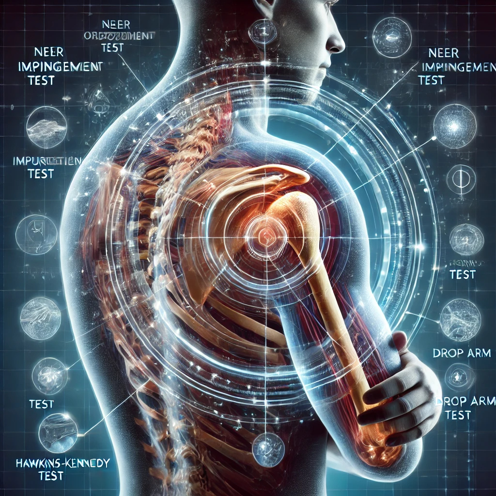
Below is a detailed list of common orthopedic examination tests for the shoulder joint. Each test includes a brief description, what it assesses, and reliability/specificity information based on available evidence.
1. Neer Impingement Test
Procedure: The patient sits or stands. The examiner stabilizes the scapula with one hand while passively flexing the arm in the scapular plane to near full elevation. Internal rotation is added.
Purpose: Detects subacromial impingement or rotator cuff irritation.
Reliability: Moderate reliability.
Specificity: Low (50–60%), as it can produce false positives in cases of other shoulder conditions.
2. Hawkins-Kennedy Test
Procedure: The patient sits with the arm flexed to 90° and the elbow bent to 90°. The examiner stabilizes the elbow and forces internal rotation.
Purpose: Assesses subacromial impingement.
Reliability: Moderate reliability.
Specificity: Low (around 50%), with better sensitivity (~80%).
3. Empty Can Test (Jobe's Test)
Procedure: The patient raises the arms to 90° in the scapular plane with thumbs pointing down. Resistance is applied downward by the examiner.
Purpose: Evaluates supraspinatus muscle strength.
Reliability: Moderate to high reliability in detecting weakness.
Specificity: Moderate (55–70%), often used in conjunction with other tests.
4. Drop Arm Test
Procedure: The examiner passively abducts the patient’s arm to 90°, then asks the patient to slowly lower it.
Purpose: Identifies supraspinatus tears or larger rotator cuff injuries.
Reliability: High reliability for large rotator cuff tears.
Specificity: High (>90%) for significant tears.
5. Speed’s Test
Procedure: The patient flexes the arm to 90° with the palm up. The examiner applies resistance against flexion.
Purpose: Assesses for biceps tendonitis or superior labral tears.
Reliability: Moderate.
Specificity: Variable (50–75%).
6. Yergason’s Test
Procedure: The patient flexes the elbow to 90° with the forearm pronated. The examiner resists supination while palpating the biceps tendon.
Purpose: Tests for biceps tendon instability or tendonitis.
Reliability: Moderate reliability.
Specificity: Moderate (50–60%).
7. Apprehension Test
Procedure: With the patient’s arm abducted to 90° and externally rotated, the examiner applies a slight anterior pressure on the humeral head.
Purpose: Detects anterior shoulder instability.
Reliability: High reliability for instability.
Specificity: High (>90%).
8. Relocation Test (Fowler Sign)
Procedure: Performed immediately after a positive apprehension test. Posterior pressure is applied to the humeral head.
Purpose: Confirms anterior instability by reducing apprehension.
Reliability: High when combined with the apprehension test.
Specificity: High (>90%).
9. Sulcus Sign
Procedure: With the patient’s arm relaxed, the examiner applies inferior traction to the humerus.
Purpose: Identifies inferior instability or multidirectional instability.
Reliability: High reliability.
Specificity: Moderate (50–80%).
10. O’Brien Test (Active Compression Test)
Procedure: The patient flexes the arm to 90° with 10–15° adduction and thumbs down. Resistance is applied downward. Repeat with the thumb up.
Purpose: Assesses labral tears (SLAP lesions).
Reliability: Moderate.
Specificity: Moderate to high (~70%).
11. Lift-Off Test
Procedure: The patient places the back of their hand on the lower back and attempts to lift it off while the examiner resists.
Purpose: Evaluates subscapularis strength.
Reliability: High reliability for subscapularis tears.
Specificity: High (>90%).
12. Belly Press Test
Procedure: The patient presses their hand against their abdomen while maintaining internal rotation.
Purpose: Detects subscapularis tears when the lift-off test cannot be performed.
Reliability: High.
Specificity: High (>90%).
13. Crank Test
Procedure: With the patient’s arm abducted to 90° and elbow flexed, the examiner applies axial compression while internally and externally rotating the arm.
Purpose: Identifies labral tears.
Reliability: Moderate.
Specificity: Moderate (~60–70%).
14. Cross-Body Adduction Test
Procedure: The examiner passively adducts the patient’s arm across the chest.
Purpose: Detects acromioclavicular (AC) joint pathology.
Reliability: Moderate.
Specificity: Moderate to high (~70%).
15. Posterior Apprehension Test
Procedure: With the patient’s arm flexed to 90° and internally rotated, the examiner applies a posterior force on the elbow.
Purpose: Tests for posterior instability.
Reliability: Moderate.
Specificity: Moderate (~60–70%).
16. Jerk Test
Procedure: The patient’s arm is abducted to 90° and internally rotated. The examiner applies an axial load while horizontally adducting the arm.
Purpose: Identifies posterior instability or posterior labral tears.
Reliability: Moderate.
Specificity: Moderate (~60–70%).
17. External Rotation Lag Sign
Procedure: The patient’s arm is passively externally rotated and then asked to maintain the position.
Purpose: Assesses infraspinatus and supraspinatus function.
Reliability: High reliability for rotator cuff tears.
Specificity: High (>90%).
Notes on Reliability and Specificity:
Reliability: Varies based on the examiner’s skill and the patient population. Standardized training improves consistency.
Specificity: Tests with high specificity are more likely to confirm a condition (true positives), while those with low specificity may yield false positives and should be corroborated with imaging or additional tests.
Below is a detailed list of common orthopedic examination tests for the knee joint, including a brief description, the condition they assess, and information about reliability and specificity.
1. Lachman Test
Procedure: The patient lies supine with the knee flexed at 20–30°. The examiner stabilizes the femur with one hand and pulls the tibia anteriorly with the other.
Purpose: Detects anterior cruciate ligament (ACL) tears.
Reliability: High (often considered the gold standard for ACL testing).
Specificity: High (>90%).
2. Anterior Drawer Test
Procedure: The patient lies supine with the knee flexed at 90°. The examiner stabilizes the foot and pulls the tibia anteriorly.
Purpose: Assesses ACL integrity.
Reliability: Moderate (less reliable than the Lachman test).
Specificity: Moderate (~50–75%).
3. Posterior Drawer Test
Procedure: Similar to the anterior drawer test but involves pushing the tibia posteriorly.
Purpose: Evaluates posterior cruciate ligament (PCL) integrity.
Reliability: High when performed correctly.
Specificity: High (>90%).
4. Varus Stress Test
Procedure: With the patient’s leg slightly flexed, the examiner applies a lateral force to the knee while stabilizing the ankle.
Purpose: Tests the lateral collateral ligament (LCL).
Reliability: Moderate to high.
Specificity: High (>90%).
5. Valgus Stress Test
Procedure: Similar to the varus stress test but involves applying a medial force to assess the medial collateral ligament (MCL).
Purpose: Detects MCL injuries.
Reliability: Moderate to high.
Specificity: High (>90%).
6. McMurray Test
Procedure: The patient lies supine. The examiner flexes the knee fully, externally or internally rotates the tibia, and extends the knee while palpating the joint line.
Purpose: Detects meniscal tears.
Reliability: Moderate (depends on examiner experience).
Specificity: Variable (50–85%).
7. Apley Compression Test
Procedure: The patient lies prone with the knee flexed to 90°. The examiner applies downward pressure on the heel while rotating the tibia.
Purpose: Identifies meniscal pathology.
Reliability: Moderate.
Specificity: Moderate to high (~70–80%).
8. Thessaly Test
Procedure: The patient stands on one leg with the knee slightly flexed (20°) and twists their body back and forth while the examiner supports their balance.
Purpose: Detects meniscal tears.
Reliability: High for diagnosing meniscal injuries.
Specificity: High (~85–90%).
9. Pivot Shift Test
Procedure: The patient lies supine with the knee extended. The examiner applies valgus stress and internal rotation to the tibia while flexing the knee.
Purpose: Assesses ACL integrity and instability.
Reliability: High but dependent on patient relaxation.
Specificity: High (~90%).
10. Posterior Sag Test
Procedure: The patient lies supine with the hip flexed to 45° and the knee flexed to 90°. The examiner observes if the tibial tuberosity “sags” posteriorly.
Purpose: Identifies PCL injuries.
Reliability: High.
Specificity: High (>95%).
11. Patellar Apprehension Test
Procedure: With the patient’s knee slightly flexed, the examiner applies lateral pressure to the patella.
Purpose: Tests for patellar instability or subluxation.
Reliability: Moderate.
Specificity: High (>85%).
12. Clark’s Test (Patellar Grind Test)
Procedure: The examiner places their hand above the patella and asks the patient to contract their quadriceps.
Purpose: Detects patellofemoral pain syndrome or chondromalacia patellae.
Reliability: Low (many false positives).
Specificity: Low (~50%).
13. Ege’s Test
Procedure: The patient squats with the feet externally rotated (medial meniscus) or internally rotated (lateral meniscus).
Purpose: Assesses for meniscal pathology.
Reliability: High for meniscal injuries.
Specificity: High (~80–90%).
14. Bounce Home Test
Procedure: The examiner passively extends the patient’s knee from a flexed position and observes for a “rubbery” end feel or incomplete extension.
Purpose: Detects meniscal tears or joint effusion.
Reliability: Moderate.
Specificity: Moderate (~70%).
15. Noble’s Compression Test
Procedure: The patient lies supine, and the examiner applies pressure to the lateral femoral epicondyle while the knee is flexed and extended.
Purpose: Identifies iliotibial band (ITB) friction syndrome.
Reliability: Moderate to high.
Specificity: High (~85%).
Below is a detailed list of common orthopedic examination tests for the hip joint, including a description, the condition being assessed, and reliability/specificity information.
1. FABER Test (Patrick’s Test)
Procedure: The patient lies supine. The examiner places the leg in a figure-four position (Flexion, ABduction, External Rotation) and applies gentle downward pressure on the knee and opposite hip.
Purpose: Assesses hip joint pathology, sacroiliac (SI) joint dysfunction, or iliopsoas tightness.
Reliability: Moderate reliability for hip or SI joint pain.
Specificity: Moderate (~60–70%).
2. FADIR Test (Flexion, ADduction, Internal Rotation)
Procedure: The patient lies supine. The examiner flexes the hip to 90°, adducts it, and internally rotates the leg.
Purpose: Detects femoroacetabular impingement (FAI) or labral pathology.
Reliability: High for FAI.
Specificity: Moderate to high (~75–90%).
3. Thomas Test
Procedure: The patient lies supine and pulls one knee to the chest while the other leg remains extended. The examiner observes whether the extended leg lifts off the table.
Purpose: Assesses hip flexor tightness (e.g., iliopsoas).
Reliability: High for identifying tightness.
Specificity: High (>90%).
4. Trendelenburg Test
Procedure: The patient stands on one leg while the examiner observes for pelvic drop on the contralateral side.
Purpose: Identifies gluteus medius weakness or hip abductor dysfunction.
Reliability: High when performed correctly.
Specificity: Moderate to high (~70–85%).
5. Ober’s Test
Procedure: The patient lies on their side with the lower leg flexed. The examiner extends and abducts the upper leg, then allows it to lower passively.
Purpose: Detects iliotibial band (ITB) tightness.
Reliability: Moderate.
Specificity: Moderate (~60–75%).
6. Scour Test (Quadrant Test)
Procedure: The patient lies supine. The examiner flexes the hip to 90° and applies axial pressure while internally and externally rotating the femur.
Purpose: Detects labral pathology, articular cartilage damage, or hip joint degeneration.
Reliability: Moderate.
Specificity: Moderate (~60–70%).
7. Ely’s Test
Procedure: The patient lies prone. The examiner passively flexes the knee and observes for hip flexion or anterior pelvic tilt.
Purpose: Assesses rectus femoris tightness.
Reliability: High for detecting muscle tightness.
Specificity: Moderate (~70%).
8. Log Roll Test
Procedure: The patient lies supine. The examiner gently rolls the leg internally and externally.
Purpose: Detects capsular laxity or intra-articular pathology such as a labral tear.
Reliability: Moderate for detecting capsular laxity.
Specificity: Moderate to high (~70–85%).
9. Stinchfield Test
Procedure: The patient lies supine and actively raises their straight leg to 30° against the examiner’s resistance.
Purpose: Assesses intra-articular hip pathology or referred pain from the lumbar spine.
Reliability: Moderate.
Specificity: High (~80–90%).
10. Piriformis Test
Procedure: The patient lies on their side with the hip flexed to 60°. The examiner stabilizes the pelvis and applies downward pressure on the knee.
Purpose: Detects piriformis syndrome or sciatic nerve irritation.
Reliability: Moderate.
Specificity: Variable (~50–75%).
11. Craig’s Test
Procedure: The patient lies prone with the knee flexed to 90°. The examiner palpates the greater trochanter and rotates the hip until the trochanter is most prominent.
Purpose: Measures femoral anteversion.
Reliability: Moderate for structural abnormalities.
Specificity: Moderate (~70%).
12. C-Sign
Procedure: The patient makes a "C" shape with their hand and places it over the lateral hip to describe pain.
Purpose: Indicates femoroacetabular impingement or labral pathology.
Reliability: High for subjective complaints of FAI.
Specificity: High (>85%).
13. Standing Flexion Test
Procedure: The patient stands while the examiner palpates the PSIS (posterior superior iliac spines) and observes movement during forward flexion.
Purpose: Assesses sacroiliac joint dysfunction.
Reliability: Low to moderate.
Specificity: Variable (~50–70%).
14. Patellar-Pubic Percussion Test
Procedure: The patient lies supine while the examiner taps the patella and listens at the pubic symphysis with a stethoscope.
Purpose: Detects femoral neck fractures.
Reliability: High for detecting fractures.
Specificity: High (>90%).
Below is a detailed list of common orthopedic examination tests for the elbow joint, including their procedure, what they assess, and information on reliability and specificity.
1. Valgus Stress Test
Procedure: The patient’s elbow is flexed to 20–30°. The examiner applies a valgus (outward) force at the elbow while stabilizing the wrist.
Purpose: Detects medial collateral ligament (MCL) or ulnar collateral ligament (UCL) injury.
Reliability: Moderate to high.
Specificity: High (~90%).
2. Varus Stress Test
Procedure: The patient’s elbow is flexed to 20–30°. The examiner applies a varus (inward) force at the elbow while stabilizing the wrist.
Purpose: Assesses the lateral collateral ligament (LCL) or radial collateral ligament (RCL).
Reliability: Moderate.
Specificity: High (~85–90%).
3. Milking Maneuver
Procedure: The patient’s elbow is flexed to 90°, and the forearm is supinated. The examiner pulls the patient’s thumb to create valgus stress at the elbow.
Purpose: Detects UCL instability.
Reliability: High for UCL injuries.
Specificity: High (>85%).
4. Moving Valgus Stress Test
Procedure: The examiner flexes the patient’s elbow to 120° while applying valgus stress, then extends the elbow to 30°, maintaining the stress.
Purpose: Identifies UCL insufficiency, especially for throwing athletes.
Reliability: High in experienced hands.
Specificity: High (~95%).
5. Cozen’s Test
Procedure: The patient makes a fist, pronates the forearm, and extends the wrist while the examiner applies resistance.
Purpose: Detects lateral epicondylitis (tennis elbow).
Reliability: Moderate.
Specificity: Moderate to high (~70–85%).
6. Mill’s Test
Procedure: The examiner palpates the lateral epicondyle while passively flexing the patient’s wrist with the elbow fully extended.
Purpose: Assesses lateral epicondylitis.
Reliability: Moderate.
Specificity: Moderate (~75%).
7. Maudsley’s Test (Third Finger Test)
Procedure: The patient resists downward pressure applied by the examiner to the third finger.
Purpose: Evaluates lateral epicondylitis by stressing the extensor digitorum.
Reliability: Moderate.
Specificity: Moderate (~70–80%).
8. Golfer’s Elbow Test
Procedure: The examiner palpates the medial epicondyle while passively extending the patient’s wrist and fingers with the elbow fully extended.
Purpose: Detects medial epicondylitis (golfer’s elbow).
Reliability: Moderate.
Specificity: High (>85%).
9. Tinel’s Sign (Elbow)
Procedure: The examiner taps over the ulnar nerve at the cubital tunnel.
Purpose: Identifies ulnar nerve entrapment or irritation.
Reliability: Moderate (sensitivity varies with nerve pathology).
Specificity: Moderate (~70%).
10. Elbow Flexion Test
Procedure: The patient fully flexes their elbow and holds the position for 3–5 minutes.
Purpose: Detects ulnar nerve compression or cubital tunnel syndrome.
Reliability: Moderate for cubital tunnel syndrome.
Specificity: Moderate (~75%).
11. Chair Lift Test
Procedure: The patient lifts a chair with their forearm pronated and elbow extended.
Purpose: Detects lateral epicondylitis.
Reliability: Moderate to high.
Specificity: Moderate (~80%).
12. Posterolateral Rotatory Instability Test
Procedure: The patient lies supine with the arm overhead. The examiner applies axial compression and valgus stress while supinating the forearm and flexing the elbow.
Purpose: Detects posterolateral rotatory instability of the elbow.
Reliability: High in cases of instability.
Specificity: High (~90%).
13. Arm Bar Test
Procedure: The patient places their arm in shoulder extension, elbow flexion, and forearm pronation while the examiner applies pressure on the elbow.
Purpose: Identifies posteromedial elbow impingement.
Reliability: Moderate.
Specificity: High (~85%).
14. Resisted Supination Test
Procedure: The examiner resists supination of the forearm while palpating the radial head.
Purpose: Detects radial head instability or biceps tendon pathology.
Reliability: Moderate.
Specificity: Moderate (~70–80%).
Below is a detailed list of common orthopedic examination tests for the ankle joint, including descriptions, the conditions they assess, and details on reliability and specificity.
1. Anterior Drawer Test (Ankle)
Procedure: The patient sits with their foot in slight plantarflexion. The examiner stabilizes the tibia with one hand and pulls the calcaneus forward with the other.
Purpose: Detects anterior talofibular ligament (ATFL) injury or instability.
Reliability: High for detecting ATFL tears.
Specificity: High (~80–95%).
2. Talar Tilt Test
Procedure: The patient’s foot is held in neutral or slight plantarflexion. The examiner inverts the calcaneus while stabilizing the tibia.
Purpose: Assesses calcaneofibular ligament (CFL) and ATFL integrity.
Reliability: Moderate to high.
Specificity: High (~85–90%).
3. Squeeze Test
Procedure: The examiner compresses the tibia and fibula together at the mid-calf level.
Purpose: Identifies syndesmosis (high ankle sprain) injuries.
Reliability: Moderate.
Specificity: High (~85%).
4. External Rotation Stress Test
Procedure: The patient sits with the knee flexed at 90°. The examiner stabilizes the tibia and externally rotates the foot while the ankle is dorsiflexed.
Purpose: Detects syndesmosis injury (high ankle sprain).
Reliability: High when combined with imaging.
Specificity: High (~90%).
5. Thompson Test
Procedure: The patient lies prone with their foot off the table. The examiner squeezes the calf and observes for plantarflexion.
Purpose: Diagnoses Achilles tendon rupture.
Reliability: High for complete ruptures.
Specificity: Very high (>95%).
6. Homan’s Sign
Procedure: The patient lies supine, and the examiner dorsiflexes the ankle while squeezing the calf.
Purpose: Historically used to detect deep vein thrombosis (DVT).
Reliability: Poor (not recommended due to low sensitivity and specificity).
Specificity: Low (~50%).
7. Windlass Test
Procedure: The patient stands on a flat surface, and the examiner dorsiflexes the great toe while observing for pain.
Purpose: Identifies plantar fasciitis.
Reliability: High when symptoms are localized.
Specificity: High (~85–90%).
8. Cotton Test (Medial Talar Glide Test)
Procedure: The examiner stabilizes the distal tibia and applies a lateral force to the talus.
Purpose: Assesses syndesmosis instability.
Reliability: Moderate.
Specificity: Moderate to high (~70–85%).
9. Subtalar Tilt Test (Eversion Stress Test)
Procedure: The examiner stabilizes the tibia and everts the calcaneus.
Purpose: Evaluates deltoid ligament integrity.
Reliability: Moderate.
Specificity: High (~85%).
10. Peroneal Tendon Subluxation Test
Procedure: The patient’s ankle is dorsiflexed and everted. The examiner palpates the lateral malleolus while the patient actively resists.
Purpose: Detects peroneal tendon instability or subluxation.
Reliability: Moderate.
Specificity: High (~85%).
11. Tinel’s Sign (Ankle)
Procedure: The examiner taps over the tarsal tunnel (posterior to the medial malleolus).
Purpose: Identifies tarsal tunnel syndrome or tibial nerve irritation.
Reliability: Moderate.
Specificity: Variable (~50–75%).
12. Calcaneal Squeeze Test
Procedure: The examiner applies pressure to the medial and lateral sides of the calcaneus.
Purpose: Diagnoses calcaneal stress fractures or heel pain syndromes.
Reliability: Moderate to high for stress fractures.
Specificity: High (~85–90%).
13. Navicular Drop Test
Procedure: The examiner measures the height of the navicular bone from the floor with the foot in subtalar neutral and then again in a relaxed position.
Purpose: Assesses for flatfoot deformity or pronation.
Reliability: Moderate.
Specificity: Variable.
14. Figure-of-Eight Measurement
Procedure: The examiner uses a tape measure to assess swelling by wrapping it in a figure-of-eight pattern around the ankle and foot.
Purpose: Quantifies ankle swelling.
Reliability: High for swelling measurement.
Specificity: Not diagnostic (used for tracking swelling).
Here is a detailed list of common orthopedic examination tests for the sacroiliac (SI) joint, including how they are performed, what they assess, and information on reliability and specificity.
1. FABER Test (Patrick’s Test)
Procedure: The patient lies supine. The leg is placed in a figure-four position (Flexion, ABduction, External Rotation), and the examiner gently presses down on the knee while stabilizing the opposite hip.
Purpose: Assesses SI joint pain or dysfunction, as well as hip or iliopsoas pathology.
Reliability: Moderate.
Specificity: Moderate (~60–75%).
2. Fortin Finger Test
Procedure: The patient points with one finger to the area of maximal pain, typically near the posterior superior iliac spine (PSIS).
Purpose: Localizes SI joint pain.
Reliability: High for identifying SI joint-related pain.
Specificity: High (~85–95%).
3. Gaenslen’s Test
Procedure: The patient lies supine with one leg hanging off the side of the table while the opposite hip and knee are flexed. The examiner applies pressure on both the flexed knee and the extended leg to stress the SI joint.
Purpose: Detects SI joint pathology or dysfunction.
Reliability: Moderate to high.
Specificity: Moderate (~70–80%).
4. Compression Test (SI Compression Test)
Procedure: The patient lies on their side, and the examiner applies downward pressure on the uppermost iliac crest.
Purpose: Compresses the SI joint to reproduce pain, indicating SI joint pathology.
Reliability: Moderate.
Specificity: Moderate (~70%).
5. Distraction Test (SI Gapping Test)
Procedure: The patient lies supine. The examiner applies outward pressure to the anterior superior iliac spines (ASIS).
Purpose: Assesses anterior SI ligament integrity or pain.
Reliability: Moderate.
Specificity: High (~80–90%).
6. Thigh Thrust Test
Procedure: The patient lies supine with one hip flexed to 90°. The examiner applies axial pressure through the femur while stabilizing the pelvis.
Purpose: Detects SI joint pain by stressing the posterior SI ligaments.
Reliability: High when combined with other SI joint tests.
Specificity: High (~80–90%).
7. Sacral Thrust Test
Procedure: The patient lies prone. The examiner applies a downward force over the sacrum while stabilizing the pelvis.
Purpose: Detects SI joint dysfunction or pain by stressing the posterior SI ligaments.
Reliability: Moderate.
Specificity: High (~80–90%).
8. Pelvic Torsion Test
Procedure: The patient lies supine. The examiner applies an alternating torsion force to the pelvis by moving the hips and knees in opposite directions.
Purpose: Identifies SI joint instability or asymmetry.
Reliability: Moderate.
Specificity: Variable (~60–75%).
9. Standing Flexion Test
Procedure: The examiner palpates the posterior superior iliac spine (PSIS) on both sides and observes their movement during forward bending.
Purpose: Assesses for SI joint dysfunction or asymmetry.
Reliability: Low to moderate.
Specificity: Low (~50–60%).
10. Stork Test (Gillet Test)
Procedure: The patient stands on one leg and flexes the opposite hip to 90°. The examiner palpates the PSIS and observes its movement.
Purpose: Detects SI joint dysfunction or restricted mobility.
Reliability: Moderate.
Specificity: Moderate (~60–75%).
11. Yeoman’s Test
Procedure: The patient lies prone. The examiner stabilizes the pelvis and extends the hip while applying downward pressure on the SI joint.
Purpose: Assesses anterior SI ligament or lumbar spine pain.
Reliability: Moderate.
Specificity: Moderate (~70%).
12. Flamingo Test
Procedure: The patient stands on one leg while the examiner observes for pain or instability.
Purpose: Detects SI joint pain or instability.
Reliability: Moderate.
Specificity: Variable (~50–70%).
13. Single-Leg Stance Test
Procedure: The patient stands on one leg while the examiner applies downward pressure on the opposite iliac crest.
Purpose: Identifies SI joint or pelvic instability.
Reliability: Moderate.
Specificity: Moderate (~70%).
Summary of SI Joint Testing:
Reliability: Tests like the FABER, Thigh Thrust, Sacral Thrust, and Distraction tests have higher reliability, especially when used together.
Specificity: A cluster of tests increases diagnostic accuracy. For example, if three or more tests in a cluster are positive, the sensitivity is ~90% and specificity is ~85% for detecting SI joint dysfunction.
Below is a detailed list of common orthopedic examination tests for the lumbar spine, including their procedures, purpose, and details about reliability and specificity.
1. Straight Leg Raise (SLR) Test
Procedure: The patient lies supine. The examiner lifts one leg with the knee extended, stopping if pain or tightness occurs. Pain in the back or along the sciatic nerve distribution (buttock, posterior thigh, calf) may indicate nerve root irritation.
Purpose: Assesses nerve root compression or irritation, commonly from lumbar disc herniation (L4–S1).
Reliability: High for detecting disc herniation.
Specificity: Moderate (~40–70%). Sensitivity is higher (~80–90%).
2. Crossed Straight Leg Raise Test
Procedure: Similar to the SLR test, but the opposite leg (non-symptomatic side) is lifted. Pain in the affected leg during this test is indicative.
Purpose: Suggests disc herniation with significant nerve root irritation.
Reliability: High for diagnosing large disc herniations.
Specificity: High (~90%).
3. Slump Test
Procedure: The patient sits upright with hands behind their back. The examiner asks the patient to slump forward, flex the cervical spine, extend one knee, and dorsiflex the foot. Pain or discomfort indicates nerve tension.
Purpose: Evaluates neural tension or lumbar nerve root impingement.
Reliability: High for neural tension.
Specificity: Moderate (~75%).
4. Schober’s Test
Procedure: The examiner marks a point 10 cm above and 5 cm below the lumbosacral junction (approximately over L5). The patient bends forward, and the distance between the marks is measured. Limited increase (<5 cm) suggests restricted lumbar spine mobility.
Purpose: Detects lumbar spine stiffness, often associated with ankylosing spondylitis.
Reliability: Moderate to high for ankylosing spondylitis.
Specificity: High (~90%).
5. Prone Instability Test
Procedure: The patient lies prone with the torso on a table and feet on the floor. The examiner applies pressure over the lumbar spine. The patient then lifts their legs off the floor, and pressure is reapplied. Reduction of pain in the second position indicates instability.
Purpose: Identifies lumbar spine instability.
Reliability: High when used by experienced clinicians.
Specificity: Moderate (~85%).
6. Quadrant Test
Procedure: The patient stands and extends, side-bends, and rotates the lumbar spine to the side of pain. The examiner applies gentle overpressure in this position.
Purpose: Detects lumbar facet joint dysfunction or foraminal compression.
Reliability: Moderate.
Specificity: Moderate (~70–80%).
7. Patrick’s Test (FABER)
Procedure: The patient lies supine, and the examiner places the leg in a figure-four position (Flexion, ABduction, External Rotation). Downward pressure is applied to the knee and opposite ASIS.
Purpose: Differentiates lumbar spine pathology from SI joint or hip issues.
Reliability: Moderate.
Specificity: Moderate (~60–75%).
8. Beevor’s Sign
Procedure: The patient lies supine and performs a partial sit-up. The examiner observes for asymmetry in the movement of the umbilicus.
Purpose: Indicates thoracic or lumbar nerve root pathology (T10–T12).
Reliability: Moderate.
Specificity: High for nerve root lesions (~85–90%).
9. Waddell’s Signs
Procedure: A set of non-organic or behavioral responses during examination, including superficial tenderness, pain on simulated testing, and inconsistent results.
Purpose: Identifies non-physiological or psychosocial contributors to pain, not specific to pathology.
Reliability: Variable.
Specificity: Low but useful as a part of a broader assessment.
10. Kemp’s Test
Procedure: The patient is seated or standing. The examiner guides the patient into lumbar extension, rotation, and lateral flexion. Pain radiating down the leg suggests radiculopathy, while localized pain may indicate facet joint dysfunction.
Purpose: Differentiates between radiculopathy and facet joint pathology.
Reliability: Moderate.
Specificity: Moderate (~70%).
11. Milgram’s Test
Procedure: The patient lies supine and lifts both legs approximately 5–10 cm off the table, holding the position for 30 seconds. Pain or inability to hold the position suggests lumbar pathology.
Purpose: Assesses intrathecal or nerve root irritation.
Reliability: Low to moderate.
Specificity: Low (~50–60%).
12. Standing Stork Test
Procedure: The patient stands on one leg and extends the lumbar spine. Pain in the low back suggests pars interarticularis stress or spondylolysis.
Purpose: Detects spondylolysis or lumbar instability.
Reliability: Moderate.
Specificity: Moderate (~75%).
13. Ely’s Test
Procedure: The patient lies prone, and the examiner passively flexes the knee. If the hip flexes simultaneously, it may indicate tight rectus femoris or femoral nerve irritation.
Purpose: Assesses lumbar nerve root irritation or tightness of the rectus femoris.
Reliability: Moderate.
Specificity: Moderate (~70%).
14. Femoral Nerve Stretch Test
Procedure: The patient lies prone. The examiner extends the hip while keeping the knee flexed. Pain in the anterior thigh indicates femoral nerve involvement.
Purpose: Detects femoral nerve root compression (L2–L4).
Reliability: Moderate to high.
Specificity: High (~85%).
Summary of Reliability and Specificity:
High Specificity Tests: Tests like the Crossed SLR, Femoral Nerve Stretch Test, and Schober’s Test are highly specific for diagnosing specific lumbar conditions.
Reliability: Tests like the SLR, Slump Test, and Thigh Thrust are reliable for identifying lumbar radiculopathy or disc herniation.
Here is a detailed overview of orthopedic examination tests for the cervical spine, including their procedures, purpose, reliability, and specificity.
1. Spurling’s Test
Procedure: The patient sits upright. The examiner applies axial compression while the neck is extended, rotated, and side-bent toward the symptomatic side. Pain radiating into the arm indicates a positive test.
Purpose: Identifies cervical radiculopathy caused by nerve root compression.
Reliability: High.
Specificity: High (~90%).
2. Cervical Distraction Test
Procedure: The patient sits or lies down. The examiner applies upward traction to the head. A reduction in radicular pain is a positive test.
Purpose: Relieves nerve root compression or foraminal narrowing.
Reliability: High.
Specificity: Moderate to high (~70–90%).
3. Sharp-Purser Test
Procedure: The patient sits upright with the head slightly flexed. The examiner stabilizes the C2 vertebra and applies gentle posterior pressure on the forehead. A clunk or reduction of symptoms indicates a positive test.
Purpose: Assesses atlantoaxial instability, often associated with transverse ligament rupture.
Reliability: Moderate.
Specificity: High (~90%).
4. Alar Ligament Test
Procedure: The patient sits or lies supine. The examiner stabilizes the C2 spinous process while passively side-bending the head. Lack of movement suggests alar ligament injury.
Purpose: Evaluates alar ligament integrity.
Reliability: Moderate.
Specificity: High (~85–90%).
5. Vertebral Artery Test
Procedure: The patient lies supine. The examiner passively extends, rotates, and side-bends the neck to each side while observing for symptoms like dizziness, nausea, or nystagmus.
Purpose: Detects vertebral artery insufficiency.
Reliability: Moderate.
Specificity: High (~85–90%), but false positives can occur.
6. Upper Limb Tension Test (ULTT)
Procedure: The patient lies supine. The examiner passively abducts the shoulder, extends the elbow, supinates the forearm, and extends the wrist and fingers. Reproduction of radicular symptoms indicates nerve tension.
Purpose: Assesses cervical radiculopathy and nerve root irritation.
Reliability: High.
Specificity: Moderate (~70–80%).
7. Jackson’s Compression Test
Procedure: The patient sits upright. The examiner applies downward pressure while the neck is rotated to one side. Radicular pain indicates nerve root compression.
Purpose: Detects cervical radiculopathy or foraminal stenosis.
Reliability: Moderate.
Specificity: High (~85–90%).
8. Adson’s Test
Procedure: The patient sits, and the examiner palpates the radial pulse while abducting and extending the shoulder. The patient extends the neck and rotates it toward the tested side while taking a deep breath. Diminished pulse indicates thoracic outlet syndrome.
Purpose: Assesses for compression of the neurovascular bundle in thoracic outlet syndrome.
Reliability: Variable.
Specificity: Moderate (~75%).
9. Roos Test (EAST Test)
Procedure: The patient holds both arms in a "goalpost" position (90° abduction and external rotation), repeatedly opening and closing their hands for 3 minutes. Symptoms like pain, weakness, or numbness indicate thoracic outlet syndrome.
Purpose: Detects vascular or neurogenic thoracic outlet syndrome.
Reliability: Moderate.
Specificity: Moderate (~70%).
10. Lhermitte’s Sign
Procedure: The patient sits upright, and the examiner passively flexes the neck. A sudden "electric shock" sensation radiating down the spine or limbs is a positive test.
Purpose: Indicates cervical spinal cord pathology (e.g., multiple sclerosis, myelopathy).
Reliability: High.
Specificity: High (~85–90%).
11. Brachial Plexus Compression Test
Procedure: The examiner applies pressure over the brachial plexus at the supraclavicular fossa. Reproduction of radicular symptoms indicates a positive test.
Purpose: Assesses for brachial plexus nerve compression.
Reliability: Moderate.
Specificity: Moderate (~70%).
12. Cervical Flexion-Rotation Test
Procedure: The patient lies supine. The examiner fully flexes the cervical spine and then rotates the head to each side. Pain or reduced range of motion suggests cervicogenic headache.
Purpose: Differentiates cervicogenic headache from other headache types.
Reliability: High.
Specificity: Moderate to high (~80%).
13. Valsalva Maneuver
Procedure: The patient holds their breath and bears down as if straining during a bowel movement. Increased pain in the neck or upper extremities indicates intrathecal pressure or disc herniation.
Purpose: Detects space-occupying lesions (e.g., herniated disc, tumor).
Reliability: Moderate.
Specificity: High (~85%).
14. Neck Flexor Endurance Test
Procedure: The patient lies supine and lifts their head off the table while maintaining the chin tucked. The examiner times how long the position can be held.
Purpose: Assesses deep cervical flexor muscle endurance.
Reliability: High.
Specificity: N/A (not a diagnostic test).
Summary of Cervical Spine Testing:
High Specificity Tests: Spurling’s Test, Cervical Distraction Test, Sharp-Purser Test, and Vertebral Artery Test are highly specific for conditions like radiculopathy, instability, or vascular insufficiency.
Cluster of Tests: Combining tests like Spurling’s, Distraction, and ULTT increases diagnostic accuracy for cervical radiculopathy.
Reliability: Tests like the ULTT and Cervical Flexion-Rotation Test are highly reliable for specific conditions.